


CASES OF THE WEEK – “Thyroid Scintigraphy & Usefulness of Spect-ct Fused Imaging in Adult Hood with Missing Thyroid Gland” by Dr ShekharShikare, Consultant & HOD, Nuclear Medicine and Dr Ram Shukla, Specialist Infectious Diseases
Thyroid Scintigraphy & Usefulness of Spect-ct Fused Imaging in Adult Hood with Missing Thyroid Gland
Abstract
Adult diagnosis of lingual thyroid is usually detected because of dysphagia, airway obstruction or dysphonia but rarely identified simply, as with our patients, because of subclinical hypothyroidism. We present two cases of missing thyroid gland in adulthood and presence of lingual thyroid, which was later confirmed on 99mTechenitum scintigraphy and precisely by SPECT-CT HYBRID imaging.
Key words- Lingual thyroid, 99mTc thyroid scan & SPECT-CT imaging.
Case 1
38 years old female with history of Polycystic ovarian syndrome, Vitamin D deficiency, dysmenorrhea and found to have subclinical hypothyroidism on investigation.
TSH 7.23 (0.35-4.94 mIU/L), FT4 12.11 (9-19 pmol/L), Anti Thyroglobulin antibody 0.61 IU/ml (less than 4.11), Anti thyroperoxidase antibodies 0.14 IU/ml (0 - 5.61) Sr. Calcium 2.26 (2.10-2.60), PTH 48.20 pg/ml (15- 68.3)
Ultrasound of thyroid- Thyroid gland very difficult to visualize.
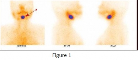
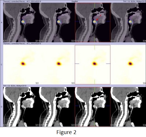
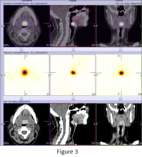
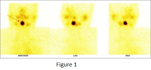
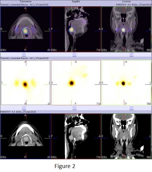
99m Technetium thyroid scan with anterior, right lateral, left lateral STATIC and SPECT-CT FUSED images of neck.
Intense focal area (1.9* 1.8 centimeter in size approximately) of isotope trapping in thyroid tissue located at the base of the tongue and better appreciated and localized in SEPCT-CT fused images & as shown with arrows (figure 2, figure3)
Thyroid bed shows no tracer uptakes (figure 1)
Case 2
29 years old male with recently diagnosed early hypothyroidism during infertility investigation.
TSH 12.9 (0.40-4.67 mIU /L), FT4 10.14 (9-19 pmol /L)
Ultrasound of thyroid showed atrophic thyroid gland. -
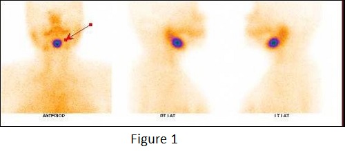
99m Technetium thyroid scan with anterior, right lateral, left lateral STATIC and SPECT-CT FUSED images of neck.
Intense focal area (1.8* 1.8 centimeter in size approximately) of isotope trapping in thyroid tissue located at the base of the tongue and better appreciated and localized in SEPCT-CT fused images & as shown with arrows (figure 2)
Thyroid bed shows no tracer uptakes (figure 1)
DISCUSSION
Nearly all (90%) of ectopic thyroid glands are found at the back of the tongue (lingual thyroid). The prevalence of lingual thyroid is one in 100,000 and occurs more often in females than males (3:1 to 7:1). Most cases of thyroid dysgenesis are sporadic.
Embryologically, the thyroid develops from the floor of the primitive pharynx and migrates anteriorly and inferiorly until it reaches its final location in the adult [3] in the final pre-tracheal position [6]. The pathogenesis of the ectopia of the thyroid tissue is not clear; however, it has been postulated that maternal antithyroid antibodies would hinder the descent of the gland during embryogenesis [1]. Thus, the ectopic thyroid tissue is the result of abnormal embryonic development and migration of the gland. It may be observed anywhere along the downward path of the gland [6].
Most patients who have lingual thyroid show no symptoms. However, there are cases where the mass can be enlarged and cause dysphagia, dysphonia, dyspnea, or a feeling of suffocation [7]. In the reported case, the difficulty in swallowing was what triggered the search for diagnosis and treatment.
The diagnosis is mainly based on clinical and imaging examinations [5]. Palpation of the neck is essential in order to check for the presence or absence of the thyroid gland in its normal position. Thyroid function tests should also be performed, but this examination may often be normal [4]. The most important medical diagnostic tool is 99m technetium thyroid scanning, computed tomography, and magnetic resonance imaging [8].
In our cases, it was possible to observe, through the SPECT-CT FUSED images, a round, well defined hyperdense nodular mass at the posterior region of the base of the tongue, which is intensely trapping up the 99m Technetium favoring presence of lingual thyroid.
The treatment for this alteration can be therapeutic or surgical and must take into account the physiological needs for thyroid hormones [5] and the severity of the symptoms [4]. The clinical control of lingual thyroid includes observation, suppressive therapy, and treatment with radioactive iodine [2].
Thyroid scintigraphy and usefulness of spect-ct fused imaging in adult hood with missing thyroid gland
Case 1
99m Technetium thyroid scan with anterior,
right lateral, left lateral STATIC and SPECT-CT FUSED images of neck.
Intense focal area (1.9* 1.8 centimeter in size approximately) of isotope trapping in thyroid tissue located at the base of the tongue and better appreciated and localized in SEPCT-CT fused images & as shown with arrows (figure 2, figure3)
Thyroid bed shows no tracer uptakes (figure 1)
Case 2
99m Technetium thyroid scan with anterior, right lateral, left lateral STATIC and SPECT-CT FUSED images of neck.
Intense focal area (1.8* 1.8 centimeter in size approximately) of isotope trapping in thyroid tissue located at the base of the tongue and better appreciated and localized in SEPCT-CT fused images & as shown with arrows (figure 2)
Thyroid bed shows no tracer uptakes (figure 1)