


CASES OF THE WEEK – “The Role of 99mtc DMSA Renal Scan in the Evaluation of Ectopic & Small Kidneys (Not Visualised on Ultrasonography)” by Dr ShekharShikare, Consultant & HOD, Nuclear Medicine
The Role of 99mtc DMSA Renal Scan in the Evaluation of Ectopic & Small Kidneys (Not Visualised on Ultrasonography) - Short Cases
A DMSA Renal scan is a radionuclide scan that uses Dimercaptosuccinic acid (DMSA) in assessing renal morphology, structure and split functions (figure 1) Radioactive technetium-99m is combined with DMSA and injected into a patient, followed by imaging with a Gamma Camera after 2-3 hours. A DMSA scan is usually static imaging,
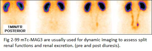
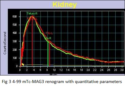
The technetium-99m DMSA binds to the proximal convoluted tubules in kidney so the excretion pattern of the kidneys cannot be assessed by this for which renal dynamic scans using radiotracers like DTPA, MAG3 are used (figures 2,3 & 4).
The major clinical indications for this investigation are small or absent kidney (renal agenesis) & Ectopic kidneys (sometimes cannot be visualized by ultrasonography of abdomen due to intestinal gas)
What is an ectopic kidney?
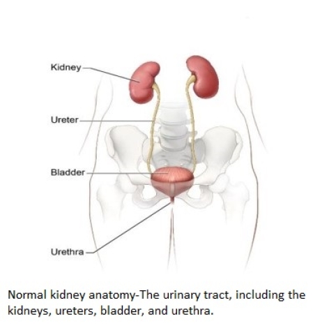
An ectopic kidney is a kidney located below, above, or on the opposite side of the kidney’s normal position in the urinary tract. The two kidneys are usually located near the middle of your back, just below your rib cage, on either side of your spine.
The urinary tract, including the kidneys, ureters, bladder, and urethra.
An ectopic kidney usually doesn’t cause any symptoms or health problems, and many people never find out that they have the condition. If an ectopic kidney is discovered, it is usually found during a fetal ultrasound—an imaging test that uses sound waves to create a picture of how a baby is developing in the womb—or during medical tests done to check for a urinary tract infection or to find the cause of abdominal pain. Rarely does a person have two ectopic kidneys.
When an ectopic kidney occurs during growth, the kidney
- stays in the pelvis near the bladder
- stops moving up too early and stays in the lower abdomen
- moves too high up in the abdomen
- crosses over the center of the body and often grows into, or joins, the other kidney, with both kidneys on the same side of the body
How common is an ectopic kidney?
Most people who have an ectopic kidney don’t have symptoms, so researchers don’t know exactly how many people have one. Some studies suggest about 1 in 1,000 people has an ectopic kidney.
Of these malformations, there exists a variety of kidney-based anatomical defects, including duplex collecting systems, crossed ectopia, and most commonly, horseshoe kidney:
Pelvic Kidney: An ectopic kidney may remain in pelvis, near the bladder (figure 5)
Crossed ectopia: Condition in which one of the kidneys crosses to the opposite end of the abdomen, and both kidneys lie on the same flank, where they can potentially be fused together. Although this condition usually has a lack of symptoms, it can result in UTIs or urinary obstructions. Patients with crossed ectopia can also experience flank pain, hematuria, or dysuria (figure 6).
Horseshoe Kidney: Most common renal abnormality found in patients with TS. Occurs during development of the fetus when the kidneys fuse together and create a horseshoe shape. Although the condition can potentially have no symptoms, the most common problems faced by a child with renal fusion include UTIs, kidney stones, and Hydronephrosis (figure 7).
Small kidney (Non visualization on ultrasonography)
Figure 8 and 9
What other health problems can an ectopic kidney cause?
An ectopic kidney usually doesn’t cause health problems, or complications, and may work normally. Most people are born with two kidneys, so if your ectopic kidney doesn’t work at all, your other kidney may be able to do the work both kidneys would have done. In rare cases, a nonfunctioning ectopic kidney must be removed. As long as the other kidney is working well, there should be no problems living with one kidney, also called a solitary kidney.
People who have an ectopic kidney are more likely to have vesicoureteral reflux (VUR). VUR is a condition in which urine flows backward from the bladder to one or both ureters, and sometimes to the kidneys. In some people, an ectopic kidney can block urine from correctly draining from the body or may be associated with VUR.
The abnormal placement of the ectopic kidney and potential problems with slow or blocked urine flow can be associated with other problems, including
Urinary tract infection. In a urinary tract with slow or blocked urine drainage or VUR, bacteria in the urine is not flushed out of the urinary tract as it normally would be, which may lead to a urinary tract infection.
Kidney stones. Kidney stones, also called urinary tract calculi, develop from minerals typically found in the urine, such as calcium and oxalate. When urine drainage is slower than normal, these minerals are more likely to build up and form kidney stones.
Trauma. An ectopic kidney in your lower abdomen or pelvis or a fused ectopic kidney may be at greater risk for damage from certain kinds of injury or trauma. Talk with a health care professional if you or your child has an ectopic kidney and wants to play contact sports or participate in other activities that may result in injury to the kidney.
TEACHING POINT
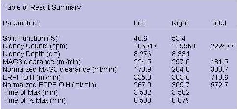
99mTc-DMSA Renal scan can be used for assessing renal morphology, structure and split functions.
Normal 99mTc-MAG3 renal scan and renogram (pre and post diuresis)
Normal 99mTc-DMSA renal scan
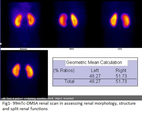
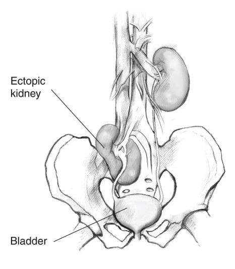
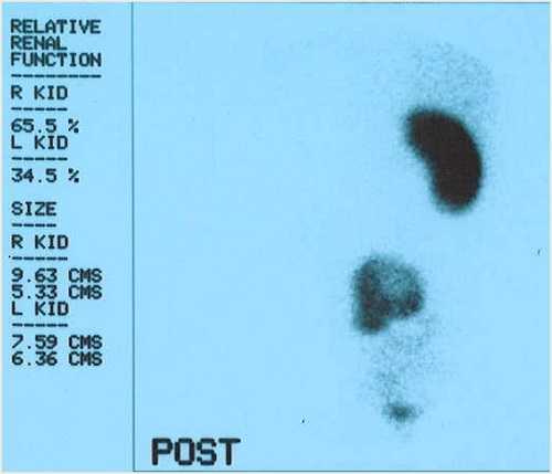
Fig5 - 99mTc-DMSA renal scan showing left ectopic pelvic kidney.
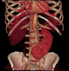
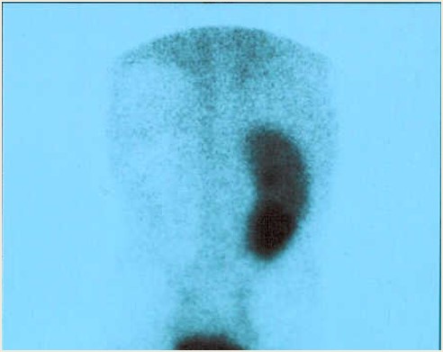
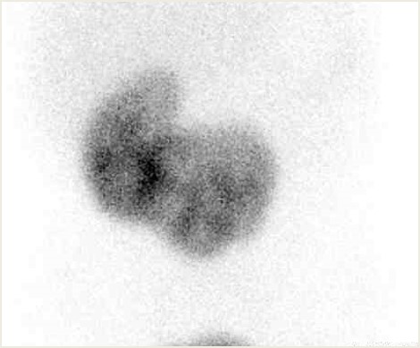
Fig 6 - 99mTc-DMSA renal scan showing left fused ectopia and right crossed fused ectopia of the kidney.
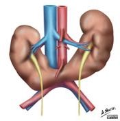
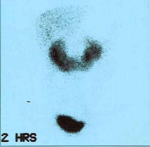
Fig 7 - 99mTc-DMSA renal scan showing horseshoe shaped kidney.