


CASES OF THE WEEK - “Imaging In Breast Cancer: Scintimammography (Smm)” by Dr ShekharShikare, Consultant & HOD, Nuclear Medicine, Al Zahra Hospital Sharjah
IMAGING IN BREAST CANCER: SCINTIMAMMOGRAPHY (SMM)
(Short interesting cases)
Although mammography remains a key imaging method for the early detection and screening of breast cancer, the overall accuracy of this test remains low. Several radiopharmaceuticals have been proposed as adjunct imaging methods to characterize breast masses by single-photon-emission computed tomography (SPECT) and positron-emission tomography (PET). Useful in characterizing indeterminate palpable masses and in the detection of axillary metastases, these techniques are insufficiently sensitive to detect subcentimetric tumor deposits. Their role in staging nodal involvement of the axillary areas therefore currently remains limited. Several enzymes and receptors have been targeted for imaging breast cancers with PET. [18F]Fluorodeoxyglucose is particularly useful in the detection and staging of recurrent breast cancer and in assessing the response to chemotherapy. Several other ligands targeting proliferative activity, protein synthesis, and hormone and cell-membrane receptors may complement this approach by providing unique information about biological characteristics of breast cancer across primary and metastatic tumor sites. Early diagnosis remains the best method of improving the odds of curing breast cancer. Among the tools currently widely available, screening mammography has been credited with an earlier diagnosis and a decreased risk of death from breast cancer. However, false negatives occur frequently, particularly when imaging post-surgical recurrence, fibrocystic breast disease and dense breast tissue in younger women. Mammography also has a low positive predictive value, and accurate second-line imaging methods are useful in some instances to reduce the number of unnecessary excisional biopsies.
Among the various imaging techniques used to assess primary or recurrent breast cancer, radionuclide imaging techniques such as planar scintigraphy, as well as SPECT-CT hybrid imaging can provide an accurate assessment of the presence and extent of disease as well as unique information about tumor biological characteristics such as the rate of proliferation and metabolic activity.
Scintimammography (SMM)
Several clinical studies in the medical literature have shown that 99mTc-MIBI, normally used for myocardial perfusion imaging, concentrates in breast cancers. This uptake is due to an increase in blood flow, number of mitochondria and cancer cell membrane hyperpolarization in the tumor and as a function of the expression of the multidrug resistance gene. 99mTc-MIBI scintimammography (SMM) has been used clinically to evaluate patients with a palpable breast abnormality when mammography is negative or indeterminate. The aggregated sensitivity and specificity of SMM in detecting a palpable primary breast cancer were, respectively, 85.2% and 86.6%. For non-palpable breast lesions, the sensitivity and specificity of SMM were 66.8% and 86.9%, respectively.
Like magnetic resonance imaging (MRI) of the breast, Nuclear Medicine may also be helpful to determine if multiple breast tumors are present. For instance, a mammogram or ultrasound (sonogram) of the breast may reveal breast cancer in one area. However, a Nuclear Medicine breast imaging test may show that the cancer is in fact multi-focal; tumors are present in several areas of the breast. Determining the extent of breast cancer with Nuclear Medicine can help indicate treatment: breast conserving surgery (lumpectomy) or breast removal (mastectomy). Mastectomy is indicated if there are multiple tumors.
Case 1
43 years old female.
Areas of suspicion in right breast upon physical examination.
Strong family history.
Mammogram findings: Extremely dense breasts. No suspicious masses or calcification.
SMM : No focal areas of abnormal tracer uptakes.
Patient did not undergo biopsy and followed by the physician.
Case 2
58 years old female.
Noticed swollen left breast three to four months ago.
sudden in onset, slowly progressive, mildly painful and redness over the skin.
No lump palpable/nipple discharge.
Similar complaints noticed in right breast two months ago.
Suspected and treated as bilateral mastitis.
USG Breast: Significantly dilated central sub-areolar ducts on either side (duct ectasia). No focal solid mass is seen. Normal sized right axillary benign looking lymph nodes with preserved fat hila noted.
Mammogram: Bilateral heterogeneous/inhomogeneous dense breast. No obvious speculated mass or suspicious clusters of micro-calcifications. The nipple retracted.
SMM: Fairly large diffuse area of abnormally increased tracer uptake seen in both the breast suggesting metabolically active abnormality. Advised Mandatory biopsy to exclude malignancy.
Bilateral US guided breast needle biopsies-Invasive duct carcinoma (NST): grade III: of both breast right and left.
Case 3
45 years old female complaining of lump and pain in her right breast. No obvious past history. Family history - Mother R.I.P with Ca breast in her mid-thirties.
O/E large lump in her right breast, lump had clearly well-defined margins and not fixed.
USG Breast: Bilateral ductal dilatation ranging between 1.7 and 6.5 mm is seen. A fairly large fibroadenoma measuring 22.4*22.3 mm is seen lateral to Right nipple shadow. One possible fibroadenoma measuring 10.2*7.8 mm is seen above the level of left nipple shadow. One possible intra-mammary lymph node 11.1.*6.5 mm
is seen superficially above and little lateral to the level of right nipple shadow. Multiple bilateral axillary lymph nodes are seen.
Mammogram: Fairly well defined hyperdense lesion > 2cms in diameter is seen lateral and cranial to the level of right nipple shadow with another small lesion seen superficially above and lateral to right nipple shadow(corresponds to larger fibroadenoma and smaller intramammary lymph node.
SMM (SCINTIMAMMOGRAPHY): Focal areas of abnormally increased tracer uptake seen in
- a) Larger one in upper quadrant of right breast.
- b) Smaller one diagonally above the larger one in right breast (Lesion a is metabolically more active than b).
- c) Upper quadrant of left breast, the tracer uptake intensity is much milder as compared to right breast abnormality (a) but looks like mirror image.
FNAC - INFILTRATING DUCT CARCINOMA (BOTH THE BREASTS)
Case 4
47 years old female. History of Total thyroidectomy (Histopathology- Papillary Ca thyroid (T1,N0,M0). Referred for 99mTc SESATMIBI whole body scan postoperative work up for residual thyroid tissue and metastasis.
Whole body 99mTc SESTAMIBI SCAN: Small residual thyroid tissue in thyroidal bed and focal area of intense uptake in Left breast(Accidentally).
Breast Biopsy- lobular breast cancer.
Case 5
61 Years old female. Strong family history of Ca breast.
Mammography: Smooth bordered possibly benign calcifications are seen scattered over both the breasts.
SMM (PLANNAR AND SPECT CT FUSED IMAGES): Focal area of abnormal MIBI tracer accumulation seen in both the breasts.
FANC BREASTS- CANCEROUS CELLS ARE SEEN IN BOTH THE BREASTS.
Case 6
56 Years old female with history of lump in right breast. Strong family history of Ca breast.
FNAC- Malignancy.
Ref for Isotope whole body bone scan for metastatic and staging work up.
99m Tc MDP Scintimammography (Done as out of interest) - Multicentric right breast lesions.
Whole body scan findings were unremarkable.
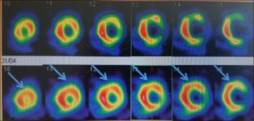
Case 7
99mTc-methoxyisobutylisonitrile (MIBI) Scinti-Mammography (SMM) Showing a typical case of breast cancer in the external upper quadrant of the left breast.
Nuclear medicine breast imaging may be appropriate for patients with:
- Equivocal, non-diagnostic or difficult to undertake x-ray mammogram.
- Dense/lumpy breast, or breast with dense masses without calcifications.
- Recurrent breast cancers.
- Strong family history of breast cancer.
- Hormone replacement therapy.
- Breast with implants.
- Palpable mass which is negative or equivocal at biopsy.
- Suspected multifocal, multicentric or bilateral disease.
- Post-operative / chemotherapy/ radiotherapy breast cancer patient.
- A lump at the surgical site after mastectomy (breast removal) since scar tissue may be difficult to distinguish from other tumors with other breast imaging exams.
Conclusion
SMM is a nuclear medicine technique, which provides valuable, additional and complimentary information to that obtained by traditional anatomical screening and assessment. SMM can provide accurate and important information in women where x-ray mammography is difficult, equivocal or non-diagnostic.Helps the clinician to achieve a more accurate diagnosis and plan the treatment more efficiently. It provides answers to questions raised by mammography and help to reduce the stress and anxiety which patients feel when undergoing tests for breast cancer by providing an increased certainty of diagnosis.
SMM should not replace mammography or ultrasonography.
NUCLEAR MEDICINE IMAGING OF MYOCARDIAL VIABILITY & ITS PRACTICAL IMPLICATION
Case 1
THALLIUM 201 IMAGES
Images of rest shows overall good viable myocardium (except in small apico-inferior portion (QGS GATED analysis- LVEF 25% EDV 257 ML, ESV 192 ML).
Case 2
THALLIUM 201 IMAGES
images of rest shows overall good viable myocardium (except relatively reduced viability in apical portion, distal half of lateral wall and distal third portion of anterior wall of the myocardium as shown in images (QGS GATED analysis-LVEF of 25%, EDV131 ml, ESV 98 ml).
Case 3
THALLIUM 201 IMAGES
Images of rest shows overall good viable myocardium
(the lateral, septal & anterior wall shows better vibaility as compared to rest of the myocardium ( QGS GATED analysis LVEF 24%, EDV 185 ml ESV 140 ml
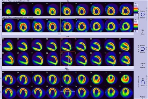
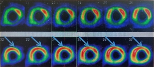
Case 4
99mTc SESTAMIBI IMAGES
Images of rest (upper line) and nitrate-enhanced rest (lower line) myocardial perfusion scintigraphy, showing improvement of perfusion in anterior (apical, medial and basal) and anterolateral (medial and basal) segments (for illustration purpose
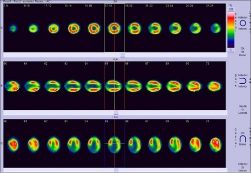
Case 5
99mTc SEASTAMIBI AND F18 FDG PET IMAGES
Myocardial perfusion scintigraphy with 99mTc-Sestamibi (upper line) and 18F-FDG PET (lower line) for assessment of myocardial viability, showing improvement in perfusion/ metabolism in anterior (apical, medial and basal), apical septal, anteroseptal (medial and basal) and inferoseptal (medial and basal) segments; “mismatch” pattern (for illustration purpose)