Special Case Surgery by Dr. Nidhi Panwar, Specialist Ophthalmology, NMC Royal Hospital Sharjah
Case – “Pneumatic Retinopexy” - A Minimal invasive technique to manage Retinal Detachment
A 56 year old female came to eye OPD with complaints of right eye nasal visual field defect since past 3-4 days and she described it as if some fly was sitting on her nose constantly.
She denied any h/o trauma, and there were no h/o floaters and flashes . She is a known hypertensive and has dyslipidemia which is controlled on medication.
On examination, her best corrected visual acuity right eye 6/6 (-4.50/-0.50*20 ) , left eye 6/6 ( -4.00/-0.50*180) , Intraocular pressure was 16 mm Hg OU. Pupils were brisk to react.
Anterior segment evaluation shows early cortical cataract both eyes.
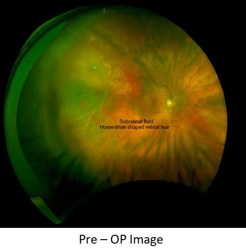
Dilated fundus evaluation of right eye reveals clear media with Disc, macula, vessels normal, Temporal peripheral retina shows a single large horse shoe tear along with surrounding subretinal fluid. Left eye media clear, disc, macula and retinal periphery normal.
In view of right eye temporal localised fresh retinal detachment along with a retinal tear with good vision, she was advised for pneumatic retinopexy, in which an expansile gas is injected in the vitreous cavity for tamponade.
Subsequently, she under cryopexy for retinal tear followed by intravitreal injection of 0.5 ml of pure SF6 gas in right eye.
Following the procedure, she was advised to maintain a strict left eye lateral position and no air travel for at least 2 weeks.
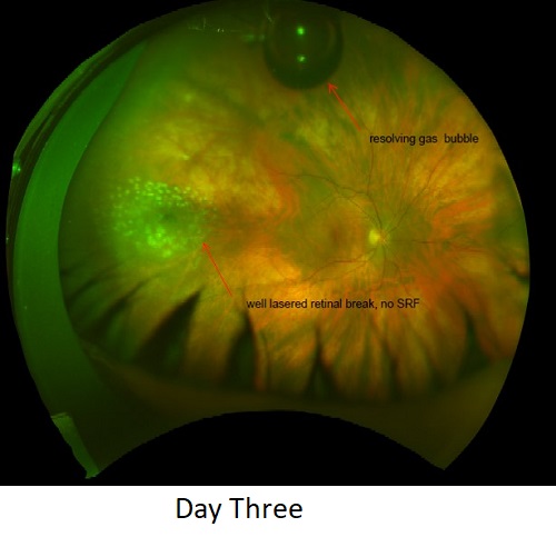
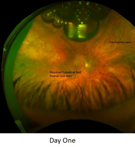
On 1st post operative day, her subretinal fluid fully disappeared and Laser could be augmented around the break. On Follow up, after 10 days of the procedure, her retina was well settled. there was almost complete resolution of the gas bubbles and no evidence of fresh tears.
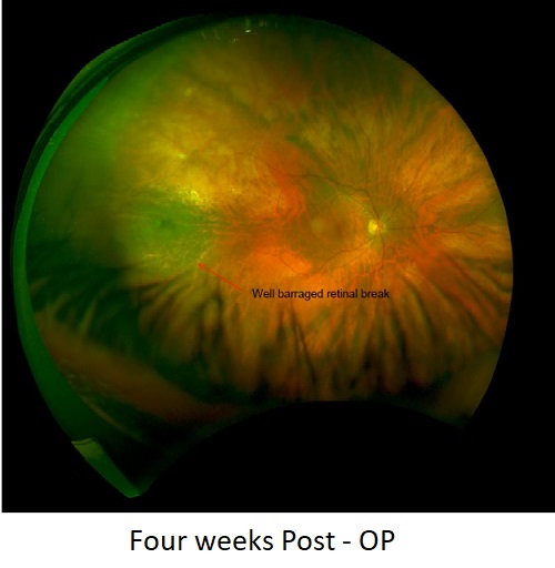
At her last follow up 4 week post procedure, her Right eye Best Corrected Visual acuity was 6/6 and IOP was 14 mm Hg without any Anti glaucoma medication, and retinal condition was stable.
Important- Rhegmatogenous Retinal detachment with an attached macula has to be treated within 1st week in order to gain maximum outcome both anatomically and functionally. Pneumatic retinopexy is very safe and effective technique in a fresh retinal detachment with retinal break above the horizontal meridian, localised with in one clock hour. . It is a minimally invasive procedure, can be done as OPD/ Day care , barely takes 10 minutes to perform, and has very minimal post operative complications.
The key is to choose the right patient and if strict criteria and right technique are applied , it can be effective in at least 90% cases avoiding the need for major vitreous retinal procedure.