


CASES OF THE WEEK – “Role of Bone Scintigraphy in the early diagnosis of Discitis” by Dr ShekharShikare, Consultant & HOD, Nuclear Medicine
Role of bone scintigraphy in the early diagnosis of Discitis
Bone scintigraphy is a valuable diagnostic procedure for discitis with earlier detection than plain radiography and similar initial detection to that of third generation computed tomography. The SPECT CT imaging has increased diagnostic confidence by indicating the involvement of the adjacent vertebral bodies rather than of the pedicles or spinous processes.
Discitis is an inflammatory disorder affecting the intervertebral disc, often following an invasive procedure but in some cases, arising a new. The clinical features, comprising fever, back pain, and constitutional symptoms, are not specific for discitis.
Both radiographic and microbiological criteria have been used to establish the diagnosis of discitis, but these criteria may not be present early in the disorder. Over the past two decades several case reports and reviews have suggested a place for scintigraphy in the detection or confirmation of discitis.
Traditionally, the diagnosis of discitis has been established by radiographic or microbiological criteria. We have shown that scintigraphy has an important role in the diagnosis, particularly after more than two weeks of symptoms. The scintigraphic criterion for discitis previously reported is a marked increase in uptake in the vertebral bodies adjacent to the affected disc. The diagnostic appearance shows that bone scintigraphy is a good indicator of discitis, comparing favorably with other imaging techniques. SPECT-CT fused imaging increases diagnostic confidence by allowing more accurate localization of the infected site.
Case
A 50-year-old lady patient presented with severe back pain along with fever. An X-ray of the spine was inconclusive.
Patient was referred for a 99mTc MDP bone scan to evaluate for spinal pathology.
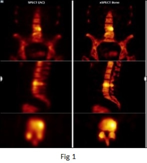
Bone SPECT-CT images shows increased uptake of the tracer in the L3-L4 lumbar vertebral end plates and intervertebral disc.
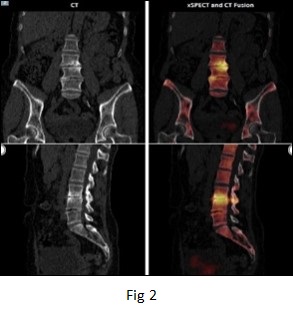
Low-dose CT shows narrowing of L3-L4 intervertebral disc space with severe erosion of the adjacent vertebral end plates along with mild sclerosis.
SPECT-CT fused data shows increased uptake within the narrowed intervertebral disc space along with uptake limited to the zone of sclerosis adjacent to the vertebral end plate erosion.
The clinical presentation of the patient, along with the SPECT-CT findings of narrowing of disc space, end plate erosion and severely increased bone metabolism within the disc space and in the adjacent vertebral end plates without major involvement of other intervertebral disc spaces, was suggestive of an intervertebral disc space infection.
The intensity of end plate tracer uptake was clearly higher than the degree of sclerosis, which is also supportive of the diagnosis of active disc space inflammation. Subsequent to the diagnosis of intervertebral disc space infection, the patient was treated with intensive antibiotic therapy.
Case
Bone SPECT shows increased uptake of the tracer in the L3-L4 lumbar vertebral end plates and intervertebral disc.
Low-dose CT shows narrowing of L3-L4 intervertebral disc space with severe erosion of the adjacent vertebral end plates along with mild sclerosis.
SPECT-CT fused data shows increased uptake within the narrowed L3-L4 intervertebral disc space along with uptake limited to the zone of sclerosis adjacent to the vertebral end plates.