CASES OF THE WEEK - “Diagnostic value of stress myocardial perfusion scintigraphy in a patient with chest pain and normal coronary angiogram” by Dr Shekhar Shikare, Consultant Nuclear Medicine & Dr Adel Abushi, Consultant Interventional Cardiology, NMC Royal Hospital Sharjah
Abstract
34 yrs old male presented with left sided chest pain radiating to the back- one episode. No h/o diabetes, hyperlipidemia, smoking, obesity, hypertention and any family history of IHD. Pulse- 91 Bpm, bp 119/84 mm of Hg, 2D Echo- WNL
STRESS MYOCARDIAL PERFUSION SCAN WITH TREADMILL EXERCISE-
Treadmill exercise with Bruce protocol- exercise time 10.29 minutes, work load achived 10.60 mets, Maxmimum heart rate 164 bpm and BP 227/79 mm of Hg, 87% of maximum age predicted heart rate. Exercise stopeed-THR achived with no symptoms.
EKG showed no significant changes.
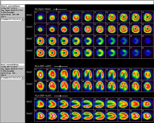
stress mpi (stress myocardial perfusion scan
MINIMAL stress induced ischemic l.v.dilatation.
MILD REVERSIBLE perfusion abnormality IN apICO-ANTERIOR AND PORTION OF ANTERIOR WALL OF THE MYOCARDIUM. MINIMAL REVERSIBLE PERFUSION ABNORMALITY IN LATERAL WALL
( DISTAL 2/3RD) AND SEPTAL WALL ( DISTAL 1.3RD), APico-inferior and inferior wall close to apex OF THE MYOCARDIUM at 87.0% of pmhr (predicted maximum heart rate) and 10.60 mets.
QUANTITATIVELY
The apico anterior and portion of anterior wall perfusion abnormality corresponds to LAD TERRITORY and is of minimal in nature ( 19%) with 19% reversibility. Rest of the perfusion abnormality as mentioned above are not quantitatively significant.The approximate total myocardium under ischemic risk is 9% with 9% reversibility.
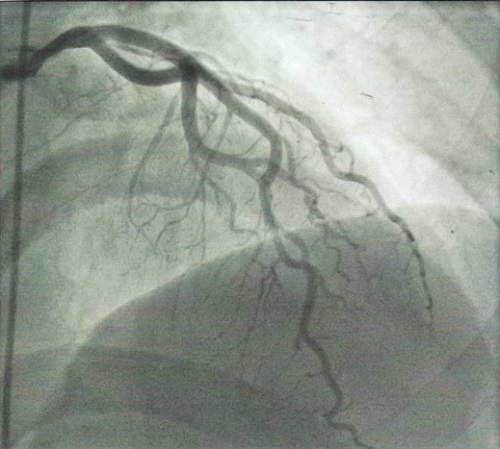
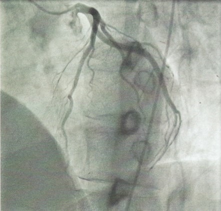
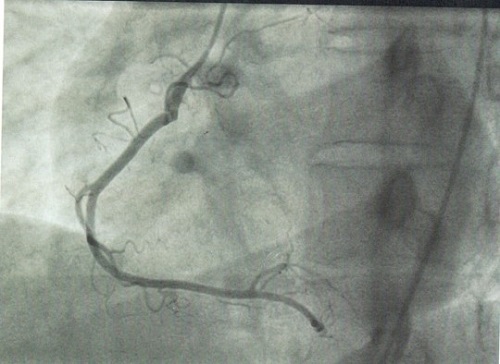
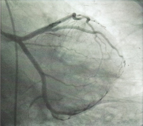
CORONARY ANGIOGRAPHY
All arteries were normal and right coronary were dominant
CONCLUSION
As per the literature- Ischemia on MPI (stress myocardial perfusion imaging) is predictive of long term adverse cardiovascular outcomes despite of normal coronary angiography ( Normal is defined here as no visible disease or luminal irregularities (< than 50%) as judged visually at coronary angiography).
There is strong correlation between the regions of the original perfusion abnormality and the ultimate coronary ischemia or revascularization.
Abnormal findings on MPI may predict a higher prevelance of coronary and peripheral vascular events than suggested by a normal coronary angiogram.
Perfusion imaging unlike angiography, closely correlated with flow reserve and reflects function of epicardial conduit arteries as well as normal capacity of the resitance vessels.
THIS CASE HIGHLIGHTS THE IMPORTANCE OF STRESS MYOCARDIAL PERFUSION SCINTIGRAPHY IRRESPECTIVE OF NORMAL CORONARY ANGIOPGRAPHY.