CASES OF THE WEEK - “Case of primary hyperparathyroidism due to an ectopic parathyroid adenoma presenting as acute pancreatitis” by Dr Shekhar Shikare, Consultant Nuclear Medicine & Dr Nishanth Sanalkumar, Consultant Endocrinology, NMC Royal Hospital Sharjah
Case of primary hyperparathyroidism due to an ectopic parathyroid adenoma presenting as acute pancreatitis.
A 52-year-old man presented with an episode of abdominal pain associated with vomiting and was diagnosed to have acute pancreatitis.
Investigation for the cause of acute pancreatitis, revealed hypercalcemia (11.8 mg/dl) low serum phosphorous and elevated parathyroid hormone (92 pg/ml). Lipid profile was normal (total cholesterol 169 mg/dL, triglyceride 125 mg/dL, LDL cholesterol 101 mg/dL, and HDL cholesterol 43 mg/dL) on atorvastatin 10 mg per day.
He denied any history of alcohol abuse and there was no family history of pancreatitis.
On the basis of biochemical parameters, a diagnosis of PHPT was made. Ultrasonography neck revealed thyroid gland to be normal in size, shape and echotexture. No focal lesions are seen. No abnormal parathyroid glands were noted.
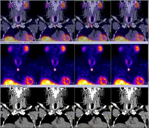
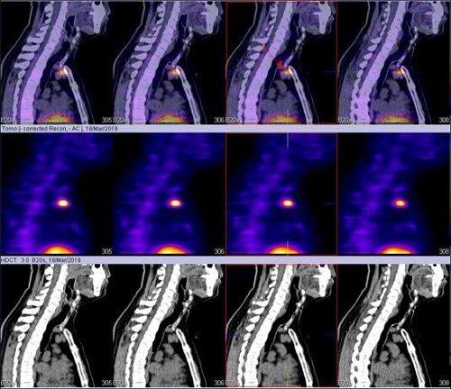
Ref for 99mTc Sestamibi Parathyroid Scan
99M TC-MIBI PARATHYROID SCINTIGRAPHY (Static early & delayed images and early SPECT CT fused images) showed focal area of abnormal MIBI tracer accumulation just behind the manubrium sterni (1.3 x 1.1 cm in size approximately) at distance of two centimeter from the thoracic inlet (Static images fig 1 and SPECT CT fused images fig 2,3,4,5). Based on this, diagnosis of retrosternal parathyroid adenoma was made.
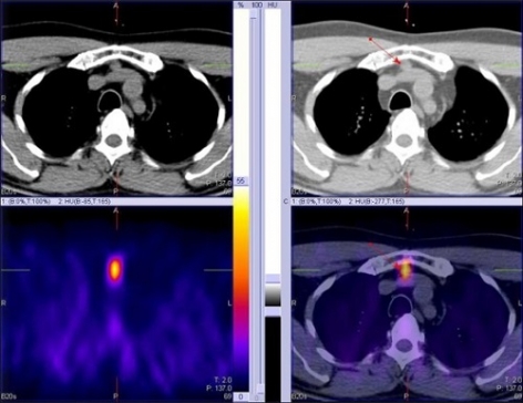
Preoperative CT with contrast revealed well circumscribed 1.4 x 1.1 cm sized, moderately & homogeneously enhancing lesion, situated about 2 cm inferior to the suprasternal notch towards right paramedian location. Patient underwent surgery through a suprasternal incision and the adenoma was removed without sternotomy. Intra operative PTH assay was done and a decline of values from 185 pg/ml to 26 pg/ml was observed. Histopathology was consistent with parathyroid adenoma. He recovered well during the post-operative period with stable serum calcium levels. He didn’t require any calcium supplements post-op.
On follow up after successful parathyroid surgery, his serum calcium was within the normal range with the last reading being 9.9 mg/dL. There has also not been any recurrence of abdominal pain or pancreatitis.