


CASES OF THE WEEK – “An improved radionuclide method for the diagnosis of Gastroesophageal reflux and aspiration in children (MILK SCAN) Background” by Dr ShekharShikare, Consultant & HOD, Nuclear Medicine
An improved radionuclide method for the diagnosis of Gastroesophageal reflux and aspiration in children (MILK SCAN) Background
Short cases
Gastroesophageal reflux disease (GERD) is the most common cause of children admissions to pediatric gastroenterology unit and affects about 30% of pediatric population. Body weight and height percentiles of children with GERD and their relationship between presence and the severity of reflux on scintigraphic images were studied.
Methods
Patients who underwent reflux scintigraphy between 2005 - 2012 were retrospectively reviewed. Among 200 patients, 49 patients were involved that their ages were ranging from 0 to 18 years old and body weight and height percentiles were recorded. Accurately 37 MBq (1 mCi) 99mTc-MAA in 100 - 150 mL of milk was ingested by the patient. Presence, number, duration and level of reflux were evaluated on the dynamic images. Presences of reflux within last ten minute were also recorded. Region-of-interests were drawn on esophagus and stomach and reflux ratio (RR) was calculated.
Results
The ratio of the presence of reflux which occurred within the last ten minutes was significantly higher in children with low body weight percentile. High-level reflux frequency was higher in these children than in normal’s. Presence of reflux which occurred within the last ten minutes was related with low body weight percentile.
Conclusions
If reflux is shown within the last ten minutes and there is high level of reflux, the clinician should be warned about possible low body weight percentile in the future and scintigraphic study should be a guide or a reference for the assessment of more effective treatment methods.
Keywords: Gastroesophageal reflux disease, Scintigraphy, Body weight
Gastroesophageal reflux disease (GERD) is the most frequently encountered esophageal disorder. GERD occurs with the passage of gastric contents into the esophagus, leading to a variety of symptoms and/or complications. The disease is the most common cause of neonatal admissions to pediatric gastroenterology unit and affects about 30% of pediatric population. Complications can be divided into two groups as esophageal and extra esophageal. The major complications are vomiting, weight loss, dysphagia, abdominal or substernal pain, esophagitis and respiratory disease. Esophageal dysfunction may also cause problems which include a series of growth and developmental retardation; therefore, implementation of early diagnosis and appropriate treatment is important.
A radionuclide study using technetium-99m-labelled milk feeding is described for the detection of gastroesophageal reflux and aspiration in pediatric age group and provide a simple, physiologic and sensitive non-invasive procedure. It has been reported that radionuclide imaging has 95%-100% sensitivity and 95%-100% specificity. Both acid and alkaline reflux can be detected and allow quantitative evaluation of reflux.
Procedure
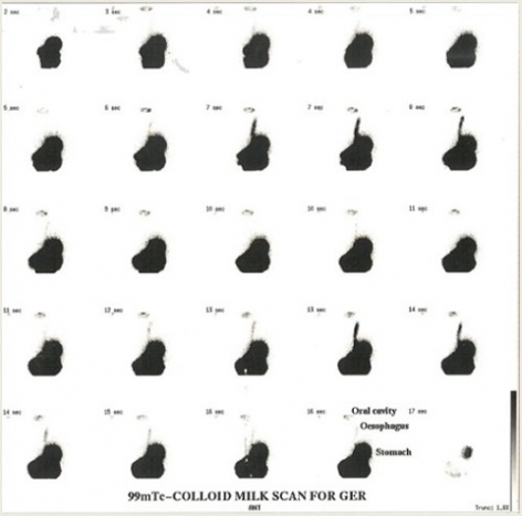
Radionuclide Reflux Scintigraphy; after 4 hours of fasting, accurately 37 MBq (1mCi) 99mTc MAA / 99mTc Colloid in 25 - 50 mL of milk was ingested by the patient (Orally or through the Ryle’s tube). Then 100 -150 mL of milk was ingested to clean up residual radioactive substance remaining in the mouth and esophagus. On supine position under the Gamma Camera, thirty-minute dynamic images are acquired & presence of reflux was evaluated. Reflux levels were determined as low (grade I), medium (grade II) and high (grade III) along with numbers of reflux episodes within 30 minutes of study were also recorded.
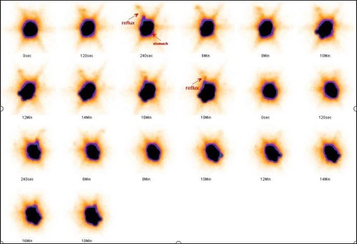
Case 1
6 days old baby, FTND, with multiple apneic spells at 6 hours of age, requiring stimulation.
99mTc-colloid labelled milk scan shows multiple episodes of tracer uptakes going up to the oral cavity region during thirty-minutes of dynamic study, suggesting high grade III GER.
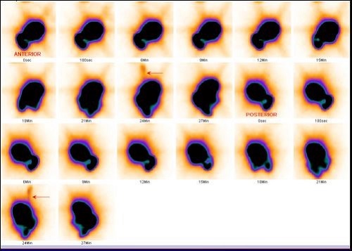
Case 2
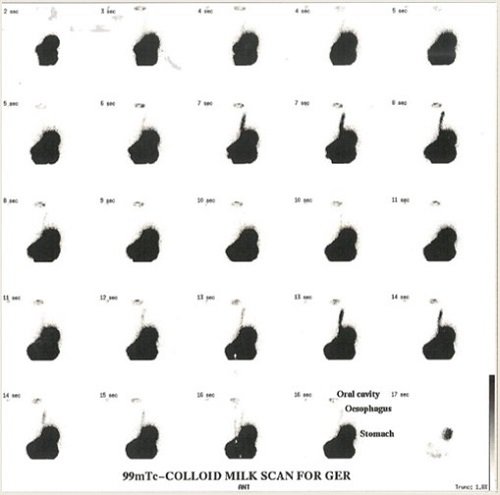
2 years old baby with history of recurrent upper respiratory tract infections and failure to thrive.
99mTc-colloid labelled milk scan shows multiple episodes of tracer uptakes going up to the oral cavity region during thirty-minutes of dynamic study, suggesting significant high-grade GER.
Case 3
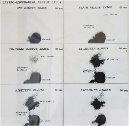
11 weeks baby with history of vomiting after feed and chocking episodes and cyanosis.
99mTc-colloid labelled milk scan shows tracer uptake visualization up to upper end of esophageal region in the initial part during thirty-minutes of dynamic study, suggesting high grade III GER.
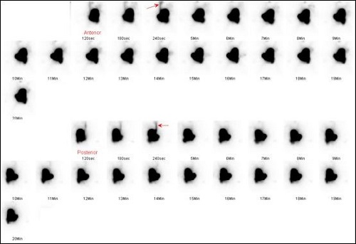
Case 4
7 days old baby with history of recurrent vomiting and cyanosis post feed.
99mTc-colloid labelled milk scan shows tracer uptake visualization up to upper end of esophageal region, twice during thirty-minutes dynamic study, suggesting grade III GER.
Case 5
6 weeks old baby with history of recurrent vomiting
99mTc-colloid labelled milk scan shows tracer uptake visualization up to upper end of esophageal region, appreciated at 24th minutes during thirty-minute dynamic study, suggesting grade III GER.
Conclusion
Milk scan is Simple, quick and more reproducible.
Sensitivity-Reflux detected in 95 to 100% symptomatic patients.
Much more sensitive than fluoroscopy and endoscopy (ST-40%) and lower esophageal sphincter pressure.
There is 90% correlation with acid pH test.
Barium swallow is an insensitive test for detecting the presence of intermittent reflux.
Although scintigraphy can detect small volumes of aspiration, the diagnosis cannot be excluded by a negative examination.
Gastro-esophageal reflux (milk scan)
Case 1
99mTc-colloid labelled milk scan shows multiple episodes of tracer uptakes going up to the oral cavity region during thirty Minutes of dynamic study, suggesting high grade III GER.
Case 2
99mTc-colloid labelled milk scan shows multiple episodes of tracer uptakes going up to the oral cavity region during thirty Minutes of dynamic study, suggesting significant high grade III GER.
Case 3
99mTc-colloid labelled milk scan shows tracer uptake visualization uptakes up to upper end of esophageal region in the initial part During thirty minutes of dymaic study, suggesting High grade III GER.
Case 4
99mTc-colloid labelled milk scan shows tracer uptake visualization up to upper end of esophageal region, twice during thirty minutes of dynamic study, suggesting grade III GER.
Case 5
99mTc-colloid labelled milk scan shows tracer uptake visualization up to upper end of esophageal region, appreciated at 24th minutes during thirty minutes of dynamic study suggesting grade III GER.