


CASE OF THE WEEK – “The role of hybrid bone SPECT/CT imaging in the work-up of the painful hip & limping patient (more symptoms with less findings)” by Dr. Shekhar Shikare, Consultant, Nuclear Medicine, Dr. Bobby Jose, HOD & Specialist, Neurosurgery & Clinical Administrator and Dr. Milind Raje, Consultant, Radiology at NMC Royal Hospital Sharjah
Introduction
Limping is an abnormal pattern of locomotion caused by pain, weakness, or deformity and many conditions of the lower limb (hip, knee, ankle, foot) can present with this common clinical presentation. Too often this symptom generates a cascade of inefficient imaging studies.
Bone hybrid SPECT/CT can be an effective problem-solving tool in patients with persistent limping when careful history taking, clinical examination, and first-line imaging modalities fail to identify the underlying cause.
HIP
The hip joint is a major weight-bearing ball-and-socket type of joint that is subjected to significant stresses during activities of daily living. Therefore, it is not surprising that more than 40% of adults over age 65 experience hip pain at some point, and 5–10% of athletes report an episode of hip pain on a yearly basis. These numbers illustrate that hip pain is indeed a very common problem.
A thorough history taking, and clinical examination are of crucial importance when assessing a patient referred for hip pain, including assessing the presence of “red flags” symptoms, as will be discussed later. The clinical examination of the adult (athletic) hip combined with the disease history will provide the clinician with a working diagnosis in approximately 80% of patients. In the 10–20% of patients in whom a working diagnosis cannot be made using this initial assessment, additional diagnostic procedures will be required
The American College of Radiology Appropriateness Criteria are heavily reliant on MR imaging, even in the setting of co-existent chronic low-back, pelvic, hip, or knee pathology. The advent of hybrid SPECT CT imaging in nuclear medicine has transformed bone scintigraphy into a comprehensive tool that not only enables whole-body skeletal assessment, in contrast to MRI, but also allows for a detailed regional evaluation using targeted SPECT/CT acquisitions. The major driver for this improvement in impact on patient management has been the increase in specificity.
Current indications for the use of bone SPECT/CT in the painful adult hip include assessment of joint replacements for loosening/infection, heterotopic ossification, impingement, stress fractures, avascular necrosis, hip pain unexplained by other imaging modalities, and ruling-out metastatic disease or systemic arthropathy. Of course, any imaging strategy should be guided by the findings on clinical examination and symptom history
Case
40 years old lady with history of sudden onset of left hip pain since last one month. It has started as on & off left hip pain, later becomes continuous & severe in nature with limping. Even at nighttime feeling the pain while turning in the bed.
X ray left hip
The visualized bone appear normal with no evidence of any fracture.
X ray lumbosacral spine
Loss of lumbar lordosis.
Reduced height of L5-S1 disc.
Facetal arthopathy at L5-S1 level.
The visualized vertebrae and rest of the bones appear normal.
MRI hip without contrast
Joint cavities are maintained. The articular surfaces are smooth. The femoral heads show normal cortical outline and cancellous marrow signal with no contusion or erosions. Small effusion is seen in the hip joint capsules. A slight thickening with focal increased signal is noted in the region of the trochanteric bursa of both the side.
Conclusion
Normal appearances of the hip joints. Minimal effusion is seen in the joint capsule and slight increased signal is noted in the region of the trochanteric bursa on both the sides.
Pelvis shows bulky uterus with multiple fibroids.
In view of the continuous symptoms, patient has been referred for the 99mTc MDP bone scintigraphy (whole body and hybrid SPECT CT) to look for the cause of the left hip pain
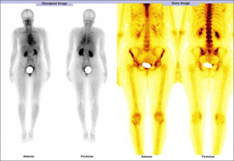
WHOLE BODY BLOOD POOL IMAGES
No obvious abnormal pooling of tracer seen in left hip region as compared to right side. Nor elsewhere on whole body images.
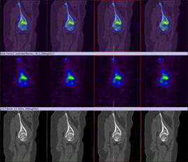
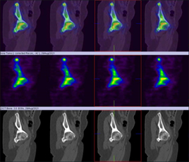
WHOLE BODY BONE, SPOT & HYBRID SPECT CT IMAGES OF HIP
It shows mildly increased tracer uptakes in left acetabular region as compared to right hip.
Rest of the skeletal show’s normal tracer uptake pattern, except mild tracer uptakes in uterus (bulky uterus and multiple fibroids on MRI).
Both the kidneys are seen.
Conclusion
Mild active abnormality in left acetabular region as compared to opposite side could be due to early osteoarthritis.
Rest of the skeletal findings are unremarkable.
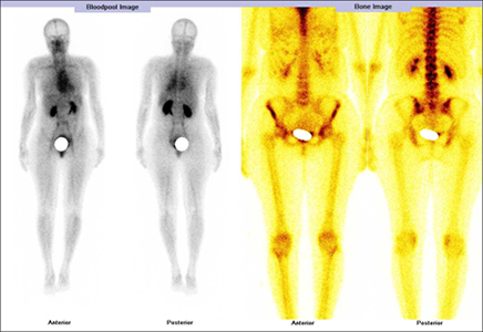
99mTc MDP whole body blood pool and bone images
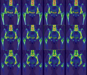
Hybrid SPECT CT bone images of hip (Coronal view)
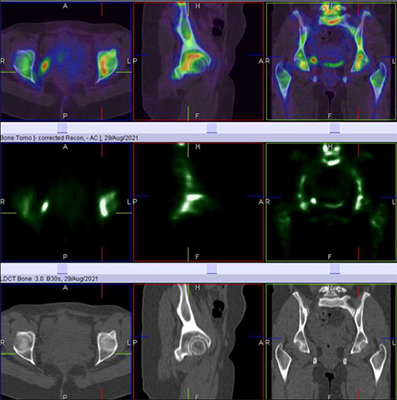
Hybrid SPECT CT bone images of both the hips
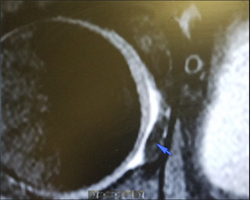
In view of the bone scan finding of the left hip, MRI images has been re-reviewed, and it shows cortical irregularity associated with focal altered signal involving medial aspect of the left femoral head along with joint effusion, which represent erosions (as shown in MRI images)
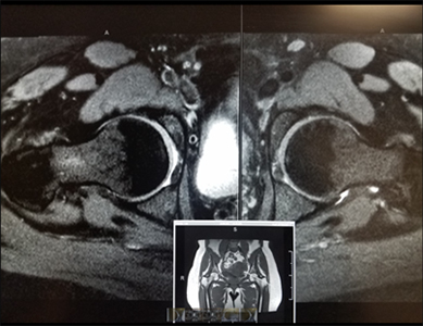
MRI images of both the hips
MRI images of right hip
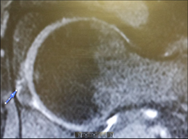
MRI images of left hip
Patient is undergoing the physiotherapy with much of the symptomatic relief.
Conclusions
Hybrid Bone SPECT/CT has shown promise to be a valuable problem-solving tool in patients with persistent pain and limping when careful history taking, clinical examination, and first-line imaging modalities fail to identify the underlying cause.as illustrated in this case.
Reference
H K Mohan 1, K Strobel 2, W van der Bruggen 3, G Gnanasegaran 4, W U Kampen 5, T Kuwert 6, T Van den Wyngaert 7 8, F Paycha 9 The role of hybrid bone SPECT/CT imaging in the work-up of the limping patient: a symptom-based and joint-oriented review. Eur J Hybrid Imaging. 2018;2(1):8. doi: 10.1186/s41824-018-0026-2. Pub 2018 Apr 23.