


CASE OF THE WEEK – “The role of hybrid bone SPECT/CT imaging in the work-up of the limping patient (Right fibular stress fracture at the syndesmosis level).” by Dr Shekhar Shikare, HOD & Consultant, Nuclear Medicine, NMC Royal Hospital Sharjah
The role of hybrid bone SPECT/CT imaging in the work-up of the limping patient (Right fibular stress fracture at the syndesmosis level).
Introduction
A vast spectrum of lower limb bone and joint disorders (hip, knee, ankle, foot) present with a common clinical presentation: limping. Too often this symptom generates an inefficient cascade of imaging studies.
The diagnostic effectiveness of bone scintigraphy using the hybrid SPECT/CT technique in relation to the diagnostic clues provided by other imaging modalities, discusses the appropriate clinical indications. Frequent lower limb bone and joint pathologies that can now be reliably diagnosed using hybrid bone SPECT/CT imaging.
Bone SPECT/CT can be an effective problem-solving tool in patients with persistent limping when careful history taking, clinical examination, and first-line imaging modalities fail to identify the underlying cause.
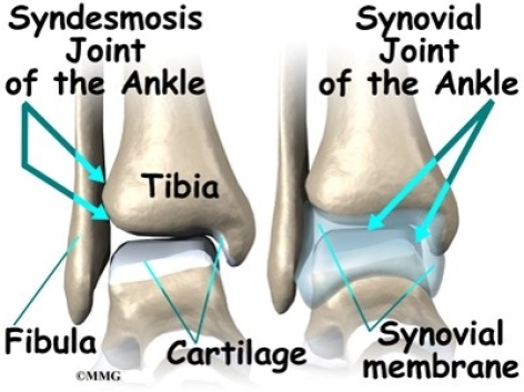
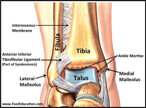
Syndesmosis
A syndesmosis is a complex fibrous joint between two bones and connected by ligaments and a strong membrane with slightly movement allowed. The distal tibiofibular syndesmosis/inferior tibiofibular joint is a syndesmotic joint. It is formed between the distal tibia (concave surface) and fibula (convex surface), with no articular capsule or synovial membrane as a fibrous joint, and attached by the interosseous ligament (IOL), the anterior-inferior tibiofibular ligament (AITFL), the posterior-inferior tibiofibular ligament (PITFL), and the transverse tibiofibular ligament (TTFL).
Syndesmosis injuries frequently occur in association with tibial or fibular fractures and MRI (3T) scanning in the plane of the syndesmotic ligaments is the investigation of choice to detect ankle syndesmosis injuries.
Case
27 years old lady with history of pain in right ankle region on walking and on movements along with limping. Known case of vitamin D deficiency.
MRI right ankle- focal sprain involving lateral fibers of inter-malleolar ligament with minimal focal sprain of the posterior talo-fibular ligament. Minimal focal sprain of tibio-talar component of the deltoid ligaments.
Ref of SPECT CT bone scan with? Stress fracture distal right fibula at syndesmosis level.
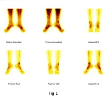
Figure 1 - 99mTc-MDP bone scan of the ankle region
Blood pool images -Focal area of abnormal pooling of tracer seen in lower end of right fibular region.
Static bone images -Focal area of increased tracer uptakes seen in lower end of right fibular bone.
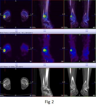
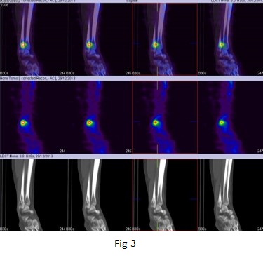
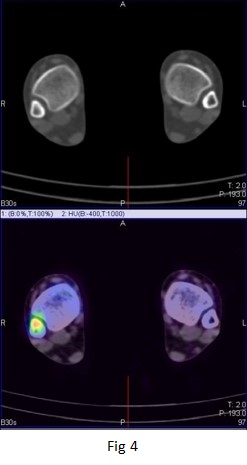
Figure 2,3, & 4 - Bone SPECT CT fused images of the right ankle
Small focally active abnormality seen at syndesmosis level of right fibular bone, favors stress fracture (figure 2 to 4)
Discussion
Limping is an abnormal pattern of locomotion caused by pain, weakness, or deformity and many conditions of the lower limb (hip, knee, ankle, foot) can present with this common clinical presentation. Too often this symptom generates a cascade of inefficient imaging studies. The introduction of hybrid SPECT/CT in nuclear medicine has drastically changed the diagnostic potential of bone scintigraphy in this setting.
Current applications of bone SPECT/CT in foot and ankle pathology typically include patients with chronic pain symptoms that are insufficiently explained by clinical findings and conventional imaging. These include locating active sites of osteoarthritis, coalitions, osteoid osteoma, occult stress fractures, tendinitis, plantar fasciitis, and impingement syndromes. The bone SPECT/CT has MRI-comparable diagnostic performance for symptomatic lesions in ankle and foot pain patients and depending on the clinical context, the technique can be proposed as second or third-line imaging modality.