


CASE OF THE WEEK – “The black Ivory vertebrae sign on Bone scintigraphy and usefulness of hybrid SPECT CT imaging” by Dr. Shekhar Shikare, HOD & Consultant, Nuclear Medicine at NMC Royal Hospital Sharjah
The ivory vertebra sign is seen at conventional radiography and refers to an increase in opacity of vertebral body that retains its size and contours, with no change in opacity and size of adjacent intervertebral discs. The increased opacity may be diffuse and homogenous and involve most or all of the vertebral body, giving it a white appearance as opposed to the normal or osteoporotic appearance of the rest of the vertebral column.
Traditionally the ivory vertebra sign has been associated with metastatic disease, especially the Carcinoma breast, Prostate and occasionally with Osteosarcoma, Carcinoid, Paget’s disease and Lymphoma, particularly Hodgkin lymphoma.
Carcinoma breast and prostate commonly results in osteoblastic metastases. A radiopaque vertebral body at one or more vertebral levels in elderly patient is most compatible with diagnosis of metastatic disease.
Solitary or more vertebral body uptake on hybrid SPECT CT Bone Scintigraphy may demonstrate “black ivory” vertebrae sign in such cases (As described by Gary S. Greene and Alan H. Maurer T) and black ivory vertebrae are correlated with their CT scan findings.
Case 1
49 years old lady with history of right breast mass, involving whole of the breast with skin involvement and no pain.
True cut biopsy of right breast mass & histopathology
Infiltrating ductal carcinoma, grade II/III. Negative for ER and PR but positive for HER2.
CONTRAST CT SCAN OF CHEST
Collapsed vertebral body of D8,
Hyperdense vertebral bodies of C7-D2, which maintain the normal size and contour, ivory vertebra.
Referred for whole body hybrid SPECT CT bone scintigraphy for the metastatic work up
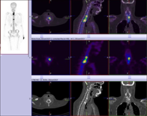
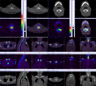
Whole body hybrid SPECT CT scintigraphy performed after intravenous (IV) administration of 14.81-mCi of 99mTc-MDP (methylene diphosphonate), revealed abnormally increased tracer uptakes in body of C7 vertebra. Left, whole body of D2 vertebra and body of D8 vertebra.
Rest of the skeletal show’s normal tracer uptake pattern.
The underlying CT images shows “IVORY” vertebra pattern in C7 and D2 vertebrae and collapsed vertebral body in D8 vertebra favoring metastatic bone disease in view of underlying disease process.
cruciate ligament. No ligamentous tear. Grade I hyperintensity seen involving posterior horn of medial meniscus.
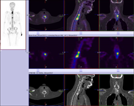
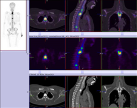
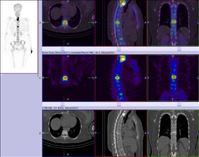
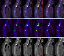
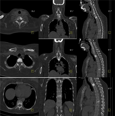
Figure 2 CT scan images shows hyperdense vertebral bodies of C7 & D2, which maintain the normal size and contour, IVORY VERTEBRAE and Collapsed D8 vertebral body.
Case 2
68-years-old gentleman is a diagnosed case of cancer prostate.
Histopathology- Prostatic adenocarcinoma, Gleason score 3+3= 6, Prognostic grade group I/V, Tumor volume 50% and PSA 15 ng/ml.
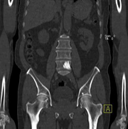
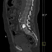
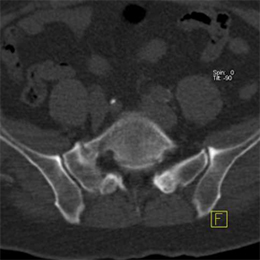
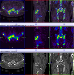
Referred for whole body Hybrid SPECT CT bone scintigraphy for the metastatic work up Whole body bone and hybrid SPECT CT scintigraphy performed after intravenous (IV) administration of 18.98 -mCi of 99mTc-MDP (methylene diphosphonate), revealed abnormally increased tracer uptakes in Body of L5 vertebra (predominantly left) extending to the left pedicle and facet joint and the underlying CT images shows “ÏVORY” vertebra pattern, which likely to represent metastasis.
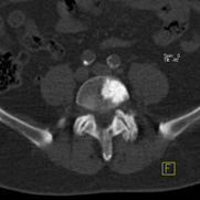
A) Body of L5 vertebra (predominantly left) extending to the left pedicle and facet joint and the underlying CT images shows “ÏVORY” vertebra pattern, which likely to represent metastasis.
A) Body of C6 & C7 approximately, both the sternoclavicular joints, Lateral ends of both the clavicles (right>left), body of S1 vertebra. Which is likely to be due degenerative pathology.
B) Right sacral bone superiorly. likely to be due to partially sacralized L5 vertebra with pseudo joint formation and showing degenerative changes.
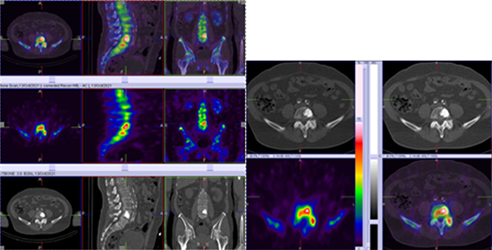
99mTc MDP Hybrid SPECT CT bone images show abnormally increased tracer uptakes in body of L5 vertebra (predominantly left), which is extending to extending to the left pedicle and facet joint, BLACK IVORY vertebra pattern, which likely to represent metastasis
99mTc MDP hybrid SPECT CT images shows abnormally increased tracer uptakes in a) Body of C6 & C7 approximately, b) Both the sternoclavicular joints c) Lateral ends of both the clavicles (right>left), Due to degenerative pathology
Teaching point
Whole-body Hybrid SPECT-CT is the optimal modality of choice for metastatic workup, including detection of extra-axial lesions, with improved sensitivity and similar specificity compared to targeted Hybrid SPECT-CT.
Reference
Gary S. Greene, MD, Mercy et all, Seminars in nuclear medicine, vol XV. No 3 (July), 1985